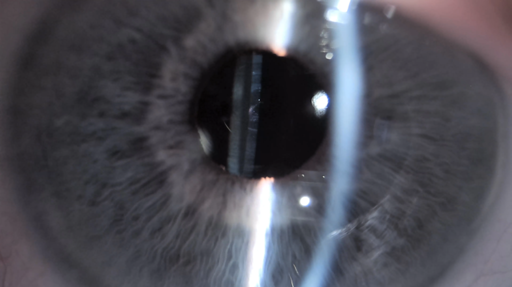


Digital image and video capture expands the applications of slit lamp microscopy. The structures of the anterior segment of the eye and any observed changes can be viewed on a screen during the examination and documented for later comparison, patient education, and professional training.
Slit lamp microscopy is a key part of examining the anterior segment of the eye. A slit lamp enables the examination of anterior eye structures using different illumination techniques and magnification levels. The examination can be used to assess, for example, the eyelids, conjunctiva, cornea, and tear film.
Traditionally, findings have been recorded in patient records in written form. A digital camera system also enables the capture of photographs and short video clips.
Image-based documentation can complement written clinical notes and facilitate the comparison of findings over time. To ensure comparability, it is important to use as consistent illumination, magnification, and imaging techniques as possible.
Images help illustrate clinical findings
One of the practical benefits of digital slit lamp microscopy is its value in patient education. The structures of the anterior segment and the findings observed during the examination can be shown directly to the patient.
For example, presenting observations of the cornea, eyelid margin, or tear film in image form can make it easier to explain the examination findings and any recommended follow-up measures.
Image material can also be used in the training of optometrists. Photographs and videos allow the same finding to be reviewed multiple times and help demonstrate examination techniques and the effects of different illumination methods.
Meibography complements the examination of meibomian glands
In the assessment of dry eye disease, evaluating the function of the meibomian glands in the eyelids is an important part of the overall examination. With appropriate imaging techniques, the gland structure can also be visualized. Meibography may provide additional information about structural changes in the glands.
Meibography visualizes gland structure, but gland function must be assessed separately as part of the clinical examination. Imaging results should always be interpreted together with symptoms, clinical findings, and other examination results. A single image alone does not determine the cause of dry eye disease and cannot replace a comprehensive clinical assessment.
Digital documentation as part of everyday clinical practice
Digital slit lamp microscopy does not replace the clinician’s expertise. Instead, it provides new opportunities for documenting findings, monitoring changes over time, and supporting patient education.
Photographs and videos make it possible to review examination findings retrospectively and to use them for professional training purposes. When storing and handling patient images, data protection requirements and the organization’s established procedures must always be followed.
Author:
Timo Juurinen, Master of Health Care, Clinical Optometry (MOptom), optometrist, Optopro Learning
Futher information:
An expert article on digital slit lamp microscopy published on Optopro.fi, including a 26-second video demonstrating imaging performed with the FREY SL-100 slit lamp and SLI-200 4K camera.