
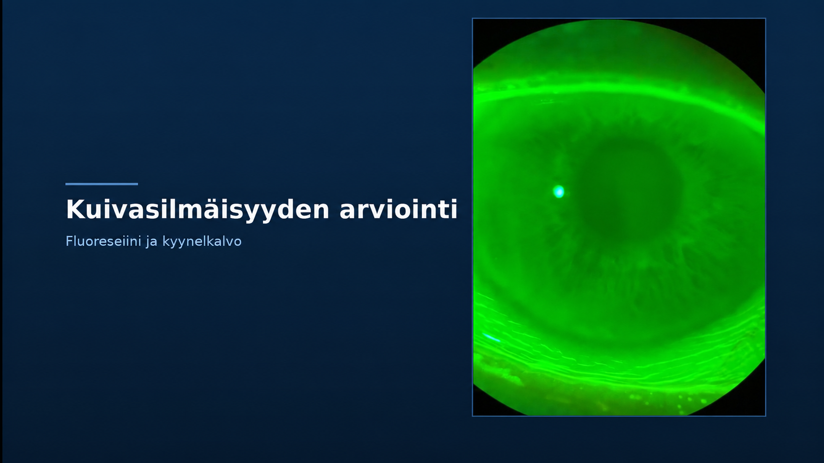


Dry eye disease is often associated with burning, a gritty sensation, excessive tearing, and fluctuating vision. However, similar symptoms may result from different abnormalities of the ocular surface and tear film. For this reason, symptoms alone are insufficient; a thorough patient history and careful examination of the ocular surface are also required. The slit lamp is one of the key tools used in this assessment.
Dry eye disease is often discussed as if it were a single condition, but in reality, it is multifactorial.
The TFOS DEWS III report, published by the Tear Film & Ocular Surface Society in 2025, defines dry eye disease as a multifactorial, symptomatic disease characterized by a loss of homeostasis of the tear film and/or ocular surface. Contributing factors may include tear film instability and hyperosmolarity, ocular surface inflammation and damage, as well as neurosensory abnormalities.
In practice, this means that dry eye should not be viewed simply as a question of whether the eye is producing too few tears.
Similar symptoms, different findings
Patients may describe their eyes as burning, dry, or gritty. Vision may fluctuate between blinks, the eyes may appear red, and sometimes excessive tearing is the most noticeable symptom.
However, symptoms alone do not reveal the underlying factors contributing to the condition. Research has shown that there can be considerable discrepancy between symptom severity and clinical findings. Dry eye symptoms and objective clinical signs do not always correlate closely.
For this reason, clinicians should form an overall assessment based on both the patient’s history and examination findings, rather than drawing conclusions from a single symptom or measurement.
The eyelid margin and meibomian glands are an important part of the picture
One of the key areas to examine is the eyelid margin.
The meibomian glands secrete lipids that contribute to the lipid layer of the tear film and help reduce tear evaporation. Meibomian gland dysfunction (MGD) is a significant factor associated with dry eye disease.
Using a slit lamp, clinicians can assess the eyelid margin, gland orifices, and meibum quality and expressibility. At the same time, other eyelid margin abnormalities may be identified and considered as part of the overall assessment.
The question is therefore not simply whether “the eye looks dry”, but rather what factors may be contributing to the observed disruption of tear film homeostasis or abnormalities of the ocular surface.
Visualizing tear film dynamics
Tear film stability is a central aspect of dry eye evaluation.
It can be assessed using a variety of methods. Non-invasive techniques allow tear film breakup to be assessed without the use of dyes. With a slit lamp and fluorescein, clinicians can assess tear film behavior as well as ocular surface staining.
During the examination, attention should be paid not only to individual numerical values but also to the overall appearance of the tear film and the location of any abnormalities.
Fluorescein may also reveal ocular surface abnormalities that might otherwise go unnoticed.
The slit lamp provides extensive information when the correct illumination technique is used
The slit lamp is a fundamental instrument for examining the anterior segment of the eye, but the quality of information obtained depends greatly on how the instrument is used.
Diffuse illumination can be used to obtain a general overview. Optical sectioning and other targeted illumination techniques help assess anterior segment structures in greater detail. Blue illumination used with fluorescein allows clinicians to assess the tear film and ocular surface staining.
The choice of examination technique therefore influences which structures and findings can be visualized during the assessment.
A systematic approach is equally important. When examinations follow the same basic protocol from one patient to another, documentation and future comparisons become easier and more reliable.
Documentation makes follow-up more meaningful
A single dry eye examination provides a snapshot of the patient’s condition at that moment.
When findings are documented systematically, changes can be compared during future visits. Information worth recording may include symptoms, tear film stability, ocular surface staining, eyelid margin findings, and observations related to meibomian gland function.
Examination conditions and blink patterns may also affect tear film measurements. For this reason, consistency in testing methods improves the reliability and comparability of follow-up data.
The goal is to identify the underlying cause behind the symptom
Symptoms indicate that something is occurring, but clinical examination helps determine what can actually be observed on the ocular surface and within the tear film.
Patient history, tear film assessment, examination of the eyelid margin and meibomian glands, and careful evaluation of the ocular surface all complement one another.
Not all dry eye presentations should be managed in the same way. Atypical, severe, or potentially sight-threatening findings, as well as those suggesting another ocular disease, require appropriate further evaluation.
For eye care professionals, understanding both the capabilities and the limitations of available examination methods is therefore essential.
Author:
Timo Juurinen, Master of Health Care, Clinical Optometry (MOptom), Optopro Learning
Timo Juurinen trains eye care professionals in slit-lamp biomicroscopy and dry eye disease. Optopro Learning has published an online course, Dry Eye Disease and Slit-Lamp Biomicroscopy in Practice, which explores the topic from the perspectives of practical examination techniques, illumination techniques, clinical findings, and documentation.
For more information and to order the course: https://leatest.fi/products/kuivasilmaisyys-ja-silmamikroskopia-kaytannossa
Sources:
Wolffsohn JS, Benítez-Del-Castillo JM, Loya-Garcia D, ym. TFOS DEWS III: Diagnostic Methodology. American Journal of Ophthalmology. 2025;279:387-450. doi:10.1016/j.ajo.2025.05.033.
Perez VL, Chen W, Craig JP, ym. TFOS DEWS III: Executive Summary. American Journal of Ophthalmology. 2026;282:135-145. doi:10.1016/j.ajo.2025.09.035.
Jones L, Craig JP, Markoulli M, ym. TFOS DEWS III: Management and Therapy. American Journal of Ophthalmology. 2025;279:289-386. doi:10.1016/j.ajo.2025.05.039.
Xiong F, Arnold BF, Lietman TM, Gonzales JA. Predictors of Discordance Between Dry Eye Symptoms and Signs. Investigative Ophthalmology & Visual Science. 2024;65(12):3.